
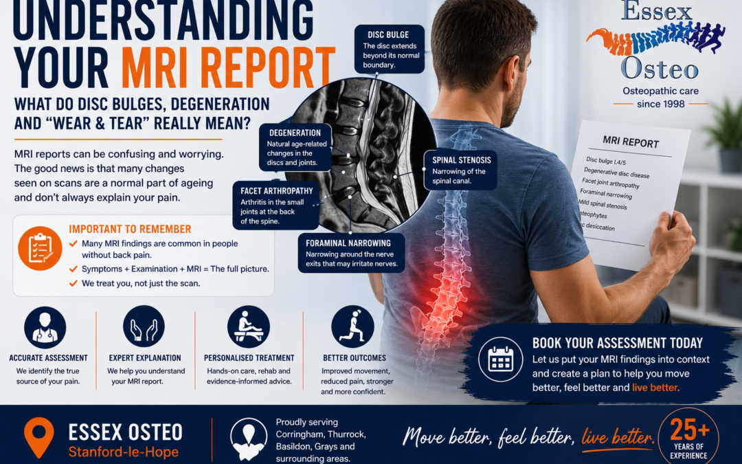
The next planned article in the series is “Understanding Your MRI Report: What Do Disc Bulges, Degeneration and Wear & Tear Really Mean?” This follows naturally from Do I Need an MRI for Back Pain? and gives us an excellent opportunity to capture people searching specific terminology from their scan reports.
Understanding Your MRI Report
What Do Disc Bulges, Degeneration and “Wear & Tear” Really Mean?
Receiving an MRI report for your lower back can sometimes create more questions than answers.
Terms such as disc degeneration, disc bulge, osteophytes, facet joint arthropathy, foraminal narrowing and spinal stenosis can sound alarming. It’s easy to read a report and assume that your spine is damaged or that the abnormalities identified must explain your pain.
But this isn’t always the case.
Many changes seen on spinal MRI scans are extremely common as we get older—and many people with exactly the same findings experience no back pain whatsoever.
At Essex Osteo in Stanford-le-Hope, we regularly see patients concerned about the wording used in their MRI reports. Understanding what these terms actually mean can provide reassurance and help ensure treatment focuses on you and your symptoms rather than simply treating an image.
Why Does My MRI Report Sound So Bad?
Radiologists have to describe everything they see.
Consequently, an MRI report might contain several findings even when none is particularly unusual for your age.
Words such as:
- Degeneration
- Degenerative change
- Disc desiccation
- Disc bulge
- Osteophytes
- Arthropathy
- Stenosis
are medical descriptions rather than predictions about how much pain you should experience.
The important question isn’t simply:
“What does the MRI show?”
It is:
“Does what we see on the MRI actually correspond with your symptoms and clinical examination?”
Abnormal MRI Doesn’t Necessarily Mean Abnormal Spine
One of the most important studies in this area reviewed MRI and CT findings from more than 3,000 people without back pain.
Age-related spinal changes were remarkably common.
For example, disc degeneration was identified in approximately:
- 37% of people aged 20
- 52% aged 30
- 68% aged 40
- 80% aged 50
- 88% aged 60
- 93% aged 70
- 96% aged 80
Disc bulges were also increasingly common with age.
This doesn’t mean MRI findings are irrelevant. Some findings can absolutely explain symptoms.
It means they need to be interpreted in the context of the individual patient.
What Does Disc Degeneration Mean?
Degenerative disc disease sounds particularly worrying, but it isn’t really a disease.
As we age, our intervertebral discs naturally change.
They may:
- Lose some water content
- Become slightly thinner
- Become less flexible
- Develop small changes within their outer fibres
An MRI may describe this as:
“Disc degeneration” or “degenerative disc disease.”
Think of it more as an age-related change than your spine wearing away.
Some people with disc degeneration experience back pain. Many don’t.
What Is Disc Desiccation?
This is another term commonly found on MRI reports.
Desiccation simply means loss of water.
Healthy spinal discs contain a high proportion of water. As we get older, they naturally become less hydrated.
On MRI this appears as a darker disc and may be reported as:
“Disc desiccation.”
Again, this is extremely common and doesn’t automatically mean the disc is causing your pain.
What Is a Disc Bulge?
A disc bulge occurs when the outer edge of an intervertebral disc extends slightly beyond its normal boundary.
Disc bulges are very common.
Importantly:
A disc bulge doesn’t necessarily mean a trapped nerve.
A bulge may be visible on MRI without touching any nerves and without causing any symptoms.
However, if the disc bulge corresponds with a patient’s symptoms and compresses or irritates a particular nerve root, it may contribute to sciatica.
Disc Bulge vs Herniated Disc
These terms are sometimes used interchangeably, but they’re not exactly the same.
A disc bulge generally involves a broader area of the disc extending beyond its normal boundary.
A disc herniation is a more focal displacement of disc material.
Herniations may be described as:
- Protrusion
- Extrusion
- Sequestration
Even these findings don’t automatically mean surgery is required.
Many disc herniations improve with conservative management, and some decrease in size over time.
What Are Osteophytes?
Osteophytes are commonly called bone spurs.
They develop around joints as part of the body’s response to age-related changes.
They are particularly common around:
- Vertebral bodies
- Facet joints
The presence of osteophytes doesn’t necessarily cause pain.
Problems are more likely if an osteophyte contributes to narrowing around a spinal nerve.
What Is Facet Joint Arthropathy?
The facet joints are small joints at the back of your spine.
Like other joints in the body, they can develop age-related osteoarthritic changes.
MRI reports may describe:
- Facet degeneration
- Facet arthropathy
- Facet hypertrophy
- Facet osteoarthritis
These changes may sometimes contribute to facet joint pain, particularly if your symptoms and examination findings correspond with that area.
What Does Foraminal Narrowing Mean?
The neural foramina are small openings on either side of the spine through which spinal nerves exit.
Changes such as:
- Disc bulging
- Osteophytes
- Facet joint enlargement
can reduce the available space.
This may be reported as:
“Neural foraminal narrowing” or “foraminal stenosis.”
Reports often grade this as mild, moderate or severe.
Foraminal narrowing becomes particularly relevant when it corresponds with symptoms such as:
- Leg pain
- Numbness
- Tingling
- Weakness
What Does Spinal Canal Stenosis Mean?
Lumbar spinal stenosis means there is reduced space within the spinal canal.
This commonly develops through a combination of:
- Disc degeneration
- Disc bulging
- Facet joint arthritis
- Thickening of spinal ligaments
Some people have considerable stenosis on MRI without major symptoms.
Others develop neurogenic claudication, where walking or prolonged standing causes leg pain, heaviness or numbness that improves when sitting or bending forwards.
We’ve covered this in greater detail in our article on Lumbar Spinal Stenosis.
What Does “Nerve Root Impingement” Mean?
This finding deserves more attention than many of the general degenerative changes.
It means something—often a disc or narrowing around the nerve—appears to contact or compress a spinal nerve root.
However, we still need to determine whether that nerve corresponds with your symptoms.
For example, if an MRI suggests compression of the L5 nerve root, we’d look for a corresponding pattern of:
- Pain
- Sensory changes
- Muscle weakness
- Reflex or functional changes
The MRI and clinical examination should tell the same story.
What Does “No Significant Neural Compression” Mean?
This is generally reassuring.
It means that although the scan may show degenerative changes or disc bulges, the radiologist hasn’t identified significant compression of the spinal nerves.
This is a common example of why reading the entire MRI report is important rather than focusing on individual words such as “degeneration.”
What Does “Modic Change” Mean?
Some MRI reports mention Modic changes.
These describe changes within the vertebral bone marrow adjacent to a spinal disc.
They are divided into different types based on their appearance on MRI.
Modic changes may be associated with low back pain in some people, but their significance varies, and their presence alone doesn’t determine your treatment.
What Is Spondylosis?
Lumbar spondylosis is an umbrella term describing age-related degenerative changes within the lumbar spine.
It may include:
- Disc degeneration
- Osteophytes
- Facet joint arthritis
- Reduced disc height
It’s essentially another way of describing common age-related changes rather than a single disease.
What Is Spondylolisthesis?
This is different from spondylosis.
Spondylolisthesis means one vertebra has moved forwards relative to the vertebra below.
Many mild spondylolistheses are stable and can be successfully managed conservatively.
We have a separate guide explaining Spondylolisthesis: Causes, Symptoms and Treatment.
Does My MRI Explain My Back Pain?
Possibly—but not necessarily.
The most useful interpretation combines three things:
Your symptoms + clinical examination + imaging findings.
For example, someone with:
- Pain travelling down the outside of the leg
- Altered sensation in an appropriate distribution
- Relevant weakness
- MRI evidence of compression of the corresponding nerve
has a much stronger clinical correlation than someone with back pain and an incidental disc bulge.
This is why a physical examination remains important even when you already have an MRI report.
Do MRI Findings Mean My Back Will Get Worse?
No.
An MRI is a snapshot of your spine at one moment in time. It doesn’t reliably predict how much pain you’ll experience in the future.
People with substantial degenerative changes may remain active and pain-free, while someone with a relatively normal-looking scan may experience significant pain.
Your spine is not simply an MRI image.
Strength, activity, sleep, general health, previous injury and many other factors influence symptoms and recovery.
Does a Disc Bulge Mean I Need Surgery?
Usually not.
Most people with disc-related back pain or sciatica are initially managed conservatively.
This may include:
- Staying active
- Appropriate pain management
- Exercise and rehabilitation
- Education
- Manual therapy as part of a treatment programme
Surgical opinion becomes more relevant when significant or progressive neurological problems develop or severe symptoms persist despite appropriate conservative management.
When Should MRI Findings Be Taken More Seriously?
Some findings require further investigation or specialist management, particularly when accompanied by concerning symptoms.
Seek urgent medical assessment if you develop:
- New bladder or bowel dysfunction
- Numbness around the saddle/genital area
- Progressive leg weakness
- Severe neurological deterioration
These can be symptoms of cauda equina syndrome and require emergency assessment.
Other concerns such as suspected fracture, infection or malignancy also require appropriate medical investigation.
See our guide: When Is Back Pain Serious? Red Flags You Should Never Ignore.
Can Osteopathy Help If My MRI Shows Degeneration?
Potentially, yes.
We don’t attempt to “reverse” normal age-related changes on an MRI.
Instead, treatment focuses on improving how you feel and function.
Depending on your assessment, treatment at Essex Osteo may include:
- Hands-on osteopathic treatment
- Joint mobilisation
- Soft tissue techniques
- Dry needling where appropriate
- Strengthening exercises
- Mobility exercises
- Individual rehabilitation
- Advice about returning to normal activity
The aim is to reduce symptoms, improve movement and help you become more confident using your back.
Bring Your MRI Report to Your Appointment
If you’ve already had an MRI, bring your report with you.
At Essex Osteo, we can discuss the terminology alongside your symptoms and clinical examination and help you understand which findings may be relevant.
This can be particularly helpful if you’ve been given your MRI results but aren’t sure what they actually mean.
Why Choose Essex Osteo?
Essex Osteo has been providing osteopathic care in Stanford-le-Hope since 1998, helping patients from Stanford-le-Hope, Corringham, Thurrock, Basildon, Grays and surrounding areas.
Our approach combines a thorough musculoskeletal assessment with evidence-informed treatment and rehabilitation.
Rather than simply treating an MRI finding, we aim to answer three questions:
What is likely causing your symptoms?
Is there anything we need to be concerned about?
What can we do to help you recover?
Frequently Asked Questions About Lumbar MRI Reports
Is disc degeneration serious?
Usually not. Disc degeneration becomes increasingly common with age and is frequently found in people without back pain.
Does a disc bulge mean I have a slipped disc?
Not necessarily. A broad disc bulge and a focal disc herniation are different imaging descriptions. Neither automatically means that the disc is causing symptoms.
Can a disc bulge go away?
Disc herniations can reduce in size over time. More general age-related disc changes may remain visible even when symptoms completely resolve.
What does mild foraminal narrowing mean?
It means the opening through which a spinal nerve exits is slightly narrower. Whether this matters depends on whether there is meaningful nerve compression and corresponding symptoms.
Does spinal stenosis always cause symptoms?
No. Some people have spinal canal narrowing on MRI without experiencing significant symptoms.
What does “degenerative change consistent with age” mean?
It generally means the radiologist has identified changes commonly seen as people get older.
Should I worry about the word “degenerative”?
Usually not. In spinal imaging, degeneration commonly describes age-related structural changes rather than a dangerous disease process.
Can you explain my MRI report at an osteopathy appointment?
Yes. An MRI can be considered alongside your medical history and physical examination to determine whether its findings are relevant to your symptoms.
Book a Back Pain Assessment in Stanford-le-Hope
If you’ve received an MRI report containing terms such as disc bulge, degeneration, stenosis, osteophytes or nerve compression and aren’t sure what they mean, a clinical assessment can help put those findings into context.
At Essex Osteo, we’ll assess your symptoms, explain relevant findings and discuss the most appropriate next steps.
Essex Osteo – Stanford-le-Hope
Providing osteopathic care since 1998.
Move better. Feel better. Live better.
Confused by your back MRI report? Learn what disc bulges, degeneration, stenosis, osteophytes and nerve compression mean. Expert guidance from Essex Osteo in Stanford-le-Hope.